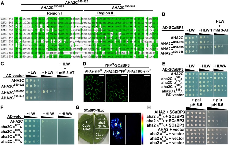
Figure 4.
SCaBP3 Interacts with PM H+-ATPase at the Region Containing the RI Domain.
(A) Amino acid sequence alignment of the C-terminal regions of PM H+-ATPases in Arabidopsis.
(B) Analysis of the interaction between SCaBP3 and AHA2 C terminus or truncated AHA2 C terminus. The C terminus was truncated into three different fragments: AH2C850-923, lacking the last 25 residues of the C terminus; AHA2C850-895, lacking the half of the AHA2 C terminus containing the RI domain; and AHA2C896-948, lacking the half of the AHA2 C terminus containing the RII domain. Cell growth was detected 3 to 5 d after transformation. Serial decimal dilutions of yeast cells were grown on synthetic complete medium without Leu and Trp (−LW, left panel), on synthetic complete medium without His, Leu, and Trp (−HLW, middle panel), and on synthetic complete medium without His, Leu, and Trp plus 3-aminotriazole (−HLW + 1 mM 3-AT, right panel).
(C) Negative control for analysis of the interaction between SCaBP3 and AHA2 C terminus or truncated AHA2 C terminus.
(D) Analysis of the interaction between SCaBP3 and AHA2 C terminus in N. benthamiana. SCaBP3, AHA2, and two truncations of AHA2 (AHA2Δ53 and AHA2Δ103) were used in BiFC experiments. AHA2Δ53, lacking the last 53 residues of AHA2; AHA2Δ103, lacking the last 103 residues of AHA2. YFP fluorescence signal was detected after 48 h of infiltration. Bars = 50 μm.
(E) Analysis of amino acids in AHA2 C terminus required for the interaction in a yeast two-hybrid analysis. The growth of the cells was detected 3 to 5 d after transformation.
(F) Negative control for analysis of amino acids in AHA2 C terminus required for the interaction(−HLWA, without His, Leu, Trp and Adenine).
(G) LCI analysis of the interaction between SCaBP3 and AHA2 or aha2 Q879A.
(H) AHA2 complementation of PM H+-ATPase activity in yeast. SCaBP3 was expressed with full-length AHA2 or different point mutations of AHA2. Cells were quintuple diluted in sterile water and grown on selective medium. AHA2, pMP1745-AHA2; aha2 G867A, pMP1745-aha2 G867A; aha2 Q879A, pMP1745-aha2 Q879A; aha2 R880A, pMP1745-aha2 R880A; gal, galactose; glu, glucose; SCaBP3, pMP1645-SCaBP3; vector, pMP1645. The growth of the cells was detected 3 to 5 d after transformation.
The experiments were performed with three biological replicates with similar results.

