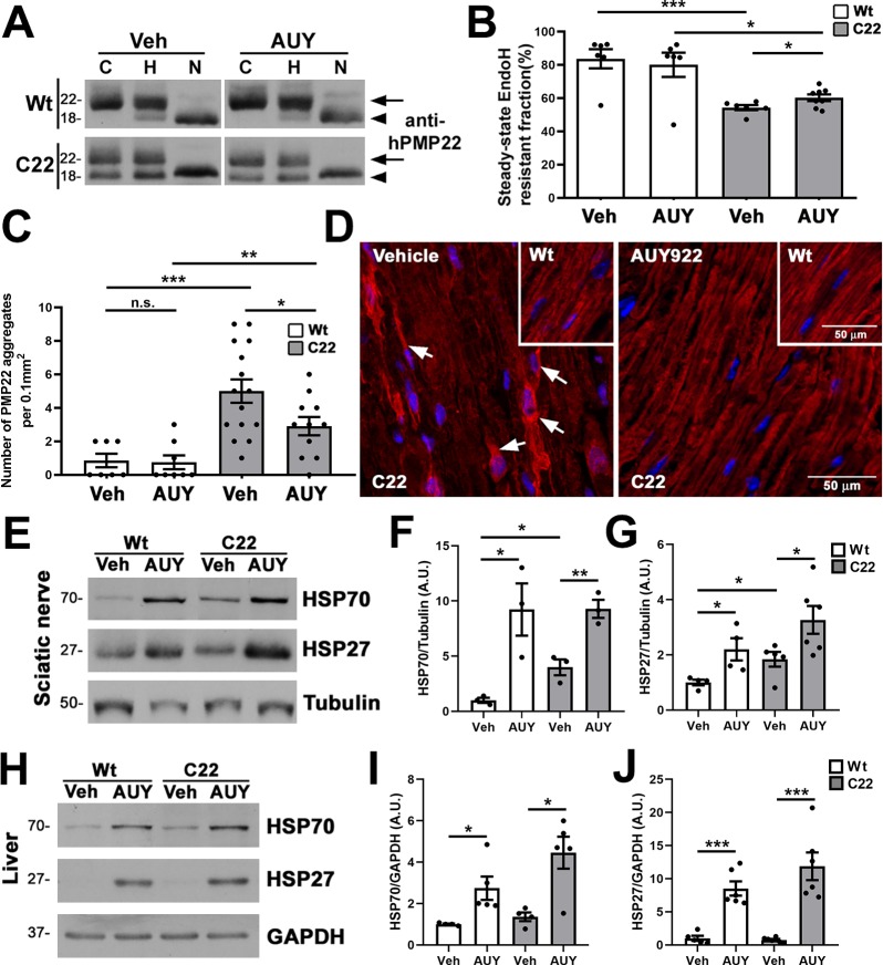
Figure 6.
Improved processing of PMP22 in AUY922-treated C22 mice. (A) Sciatic nerve lysates (5 μg/lane) were treated with either EndoH (column H) or PNGaseF (column N) and probed with antihuman PMP22 antibodies. No enzyme samples served as controls (column C). EndoH-resistant (arrows) and EndoH-sensitive (arrowheads) PMP22 fractions are marked. (B) Quantification of EndoH-resistant PMP22 fractions in sciatic nerves. (C) PMP22-positive aggregates per microscopic field (0.1 mm2) were counted in longitudinal sections of sciatic nerves. (D) Representative images of anti-PMP22 antibody stained (red) nerve sections from Wt (insets) and C22 mice are shown. Arrows mark PMP22-positive aggregates. Hoechst dye (blue) was used to visualize the nuclei. The scale bars are as shown. (E) Steady-state levels of HSP70 and HSP27 in vehicle (Veh)- and AUY922 (AUY)-treated nerve lysates (30 μg/lane) were quantified from (F, G) independent Western blots. (H) Whole liver lysates (30 μg/lane) were processed for (I, J) HSP70 and HSP27 quantification. (E–J) GAPDH or tubulin served as a loading control. Molecular mass on left in kDa. (B, C, F, G, I, J) n = 3–8 mice per group and plotted as means ± SEM; ***P < 0.001; ***P < 0.01; *P < 0.05; n.s., nonsignificant; two-tailed unpaired Student’s t-test.

