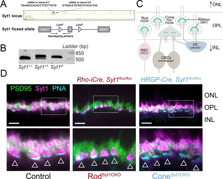
Figure 1. Syt1 was conditionally deleted from rods and cones in RodSyt1CKO and ConeSyt1CKO retinas, respectively.
(A) Top: Syt1 locus showing crRNA sequences used for inserting LoxP sites flanking exon 6; ‘/” in the amino acid sequence indicates the nucleotide positions where LoxP sites were inserted. Bottom: schematic of the Syt1flox allele showing location of genotyping primers and LoxP sites. (B) 5’ LoxP PCR of the Syt1 allele from WT (Syt1+/+) and Syt1 floxed mice (Syt1+/f: heterozygous, Syt1f/f: homozygous). Expected band sizes are 484 bp for the WT allele and 524 bp for the floxed allele. (C) Diagram illustrating fluorescent labels used for different cell types. Rod and cone terminals can be labeled with antibodies to PSD95. The base of cone terminals can be labeled with fluorescently-conjugated peanut agglutinin (PNA). Rod and cone ribbons were labeled with antibodies to CtBP2. Horizontal cells (HCs), rod bipolar cells (RBCs), and cone bipolar cells (CBCs) were labeled with antibodies to calbindin, PKCα, and secretagogin, respectively. (D) Images of control, RodSyt1CKO, and ConeSyt1CKO retinas labeled with PNA (cyan) to mark cone terminals as well as antibodies to PSD95 (green) and Syt1 (magenta). Bottom images show magnified regions outlined in the top images. Arrowheads indicate cone terminals. Scale bars = 10 µm. ONL: outer nuclear layer, INL: inner nuclear layer.

