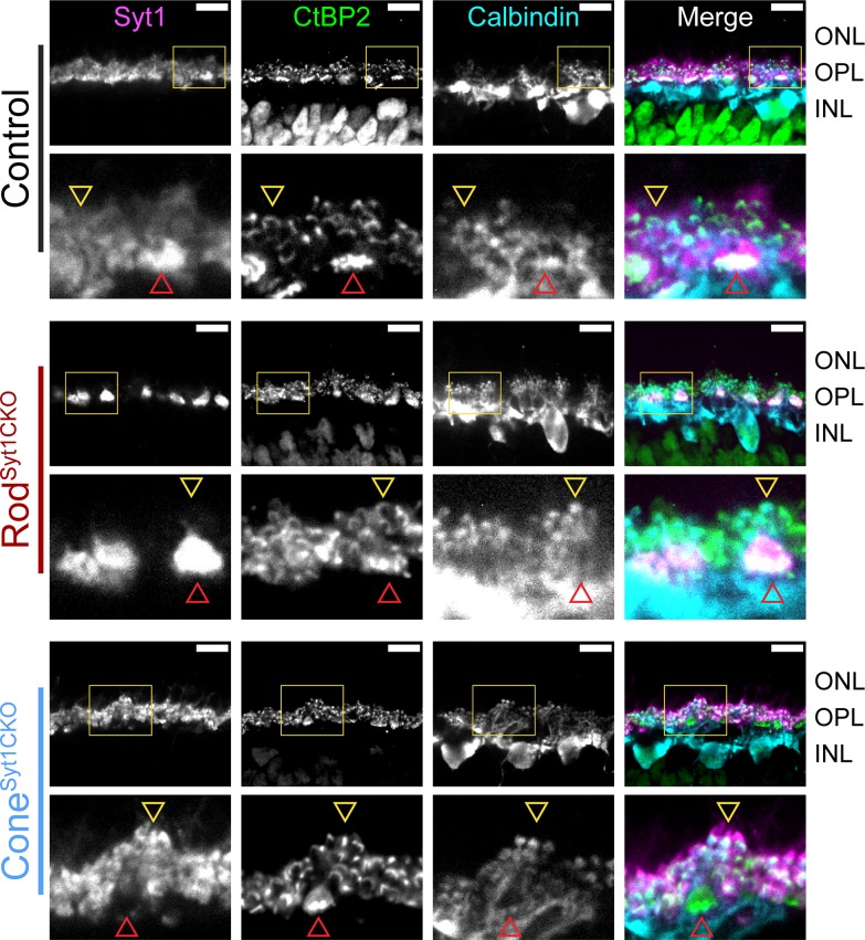
Figure 10. Horizontal cell dendrites maintain contact with Syt1-deficient rod and cone terminals in the OPL.
Images of control, RodSyt1CKO, and ConeSyt1CKO retinas labeled with antibodies to Syt1 (magenta), CtBP2 (ribbons, green), and calbindin (horizontal cells, cyan). The top row of images for each genotype contain yellow boxes that indicate the boundaries of the high magnification images below. Red arrowheads point to representative cone terminals, yellow arrowheads point to representative rod terminals. Scale bars = 10 µm.

