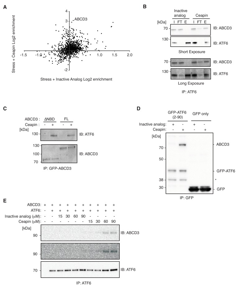
Figure 6. Ceapin drives ATF6α-ABCD3 interaction in cells and in vitro.
(A and B) Proteomic analysis and immunoblot (IB) of anti-FLAG affinity purification from 3xFLAG-ATF6α HEK293 cells treated with stress (100 nM Tg) and inactive Ceapin-A5 analog (6 μM) or active Ceapin-A7 (6 μM) with two replicates for each treatment condition. The proteins identified with affinity-purified FLAG-ATF6 treated with ER stress and Ceapin-A5 or Ceapin-A7 are listed in Figure 6—source data 1. SQSTM1 KD (*) was the top second hit in proteomics, however, SQSTM1 KD in the K562 ATF6 reporter cell line did not render cells resistant to Ceapin treatment and retained a similar response to negative control cells. I, input; FT, flow-through; E, elution. (C) Immunoprecipitation of full-length GFP-ABCD3 and GFP-ABCD3ΔNBD from cells treated with DMSO or Ceapin-A7. (D) Detergent solubilized GFP-ATF6α(2-90) or GFP-only cell lysates were incubated with Ceapin-A7 or inactive analog Ceapin-A5 and affinity purified with anti-GFP. (*) Indicates a degradation product. (E) Purified ATF6α-MBP and ABCD3-GFP were incubated with inactive Ceapin-A5 or active Ceapin-A7 and affinity purified with anti-MBP antibody.

