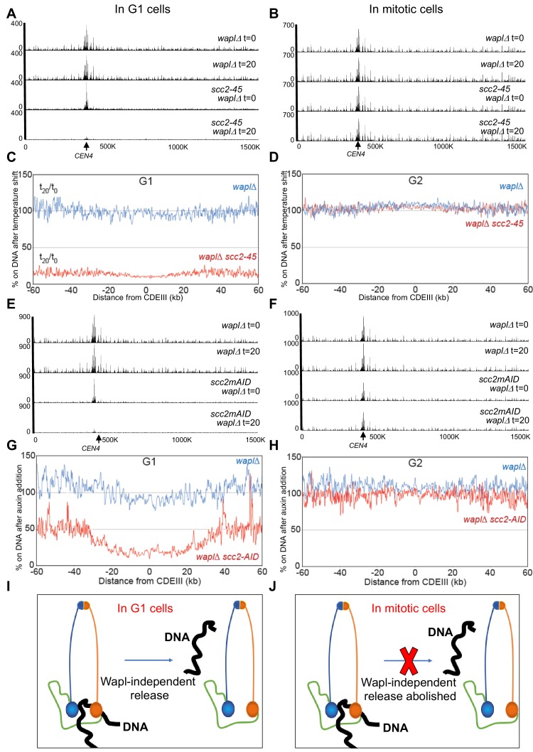
Figure 2. The Wapl-independent activity that releases cohesin from chromosomes is active only in G1 cells.
(A) waplΔ (K22296) and waplΔ scc2-45 (K22294) strains were arrested in late G1 at 25°C and subjected to temperature shift to 37°C for 20 min. 0- and 20 min samples were analysed by calibrated ChIP-sequencing (Scc1-PK6) as detailed in Materials and Methods. Cohesin ChIP profiles along chromosomes four is shown. Also see Figure 1—figure supplement 1D. (B) waplΔ (K22296) and waplΔ scc2-45 (K22294) strains were arrested in G2 with nocadazole at 25°C and subjected to temperature shift to 37°C for 20 min. 0- and 20 min samples were analysed by calibrated ChIP-sequencing (Scc1-PK6). Cohesin ChIP profiles along chromosomes four is shown. Also see Figure 1—figure supplement 1E. (C) Data form (A) is plotted to show the ratio of average cohesin levels 60 kb on either side of all 16 centromeres before and 20 min after the temperature shift. (D) Data form (B) is plotted to show the ratio of average cohesin levels 60 kb on either side of all 16 centromeres before and 20 min after the temperature shift. (E and F) waplΔ (K20891) and scc2-3XmAID waplΔ (K26831) were arrested in either late G1 or G2 and treated with auxin (IAA) for 60 min (to degrade Scc2) and subjected to Cal-ChIP-Seq. Samples drawn before (0 min) and after (60 min) auxin addition were analysed by calibrated ChIP-sequencing (Scc1-PK6). Cohesin ChIP profiles along chromosomes four is shown. Also see Figure 1—figure supplement 1H and I. (G and H) Data form (E and F respectively) are plotted to show the ratio of average cohesin levels 60 kb on either side of all 16 centromeres before and 60 min after auxin addition (I and J) The Wapl-independent activity that releases cohesin from DNA is active only in G1 (I) and not in mitotic cells (J).

