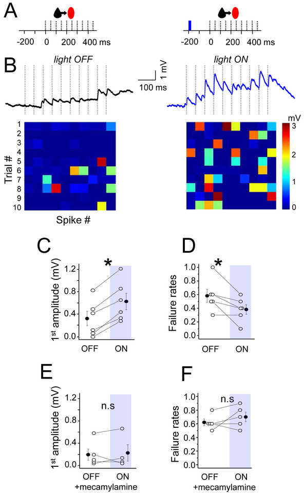
Figure 4. Nicotinic receptors enhance EPSP efficacy at Pyr-SST connections.
(A) Schematic of the stimulation protocol. 1-single blue light (10 ms) was delivered 200 ms prior to the presynaptic spike train. (B) The averaged trace of EPSP under baseline/light OFF and light ON conditions. Heatmaps at bottom show response amplitude for both conditions as described in previous figures. (C) Mean EPSP amplitude in response to the first spike in the train, for both conditions. (D) Mean failure rate after the first spike, for both conditions. (E) and (F) The same as (C) and (D) but in the presence of the nicotinic receptors antagonist (mecamylamine). For all, open circles represent individual cell measurements and filled circles represent all-cell mean± SEM, respectively (C-F). See also Figure S1 and S2.

