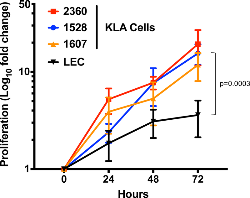
Fig. 3.

Proliferation of cells from KLA patients and human lymphatic endothelial cells (LEC). Cells were cultured in 20% FBS in EGM-2MV media on fibronectin-coated 96 well plates. Cell number was assessed at 0, 24, 48 and 72 hours using SRB assay. Cell numbers are expressed as Log10 of fold change in cell number compared to time 0. Data represents the results from 3 independent experiments.
