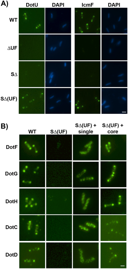
Fig 4. DotU and IcmF localize to the bacterial poles in the absence of the Legionella T4SS.
(A) DotU and IcmF localization was assayed in the wild-type strain Lp02 (WT), ΔdotU ΔicmF mutant strain (JV1181), the super dot/icm deletion strain (SΔ, JV4044) and the SΔ strain expressing dotU and icmF from the chromosome (SΔ(UF)), JV5319). Shown are Dot staining (left) and DNA stained with DAPI (right) for each set. B) Localization of Dot proteins was assayed in the wild-type strain (WT), the SΔ strain encoding dotU and icmF (SΔ(UF)), the SΔ(UF) strain expressing individual components of the core-transmembrane subcomplex (SΔ(UF) + single), and the SΔ(UF) strain expressing all five core components (SΔ(UF) + core). Antibodies used for immunofluorescence are indicated to the left of the panels. Representative images are shown from three independent experiments. Scale bar 2 μm (A,B).

