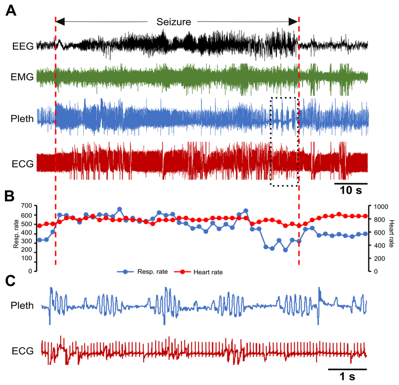
Figure 6. Spontaneous seizure in a Kcna1–/– mouse with Cheyne-Stokes respiration and no effect on heart rate.
(A) Recording of simultaneous EEG-EMG-Pleth-ECG activity during a spontaneous stage 3 seizure in a 39-day old Kcna1–/– mouse. Onset and termination of the seizure are indicated by dotted red lines. (B) Respiratory and heart rates (per min) corresponding to the recording in (A). Each dot in the plot represents the 2-s mean value. (C) Expanded traces of plethysmography and ECG for the time indicated by the boxed area showing Cheyne-Stokes respiration with no effect on the heart rate. Some movement artifacts are present in the ECG signals; therefore, scoring of heartbeats was done manually as needed. ECG, electrocardiography; EEG, electroencephalography; EMG, electromyography; Pleth, plethysmography.

