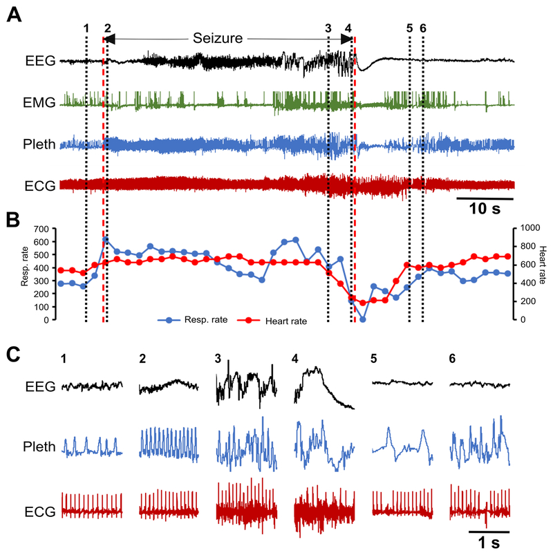
Figure 7. Spontaneous seizure in a Kcna1–/– mouse with respiratory dysfunction preceding cardiac abnormalities.
(A) Recording of simultaneous EEG-EMG-Pleth-ECG activity during a spontaneous stage 5 seizure in a 35-day old Kcna1–/– mouse. Onset and termination of the seizure are indicated by dotted red lines. (B) Respiratory and heart rates (per min) corresponding to the recording in (A). Each dot in the plot represents the 2-s mean value. (C) Expanded traces of EEG, Pleth, and ECG at the times indicated by the black dotted lines numbered 1 to 6 in (A). The numbers mark the following events: 1, pre-ictal phase; 2, hyperventilation with unchanged heart rate shortly after the onset of the seizure; 3, ictal ataxic breathing with unchanged heart rate; 4, ataxic breathing with significant bradycardia shortly before seizure termination; 5, post-ictal EEG suppression with bradypnea and a sinus exit block; 6, post-ictal ataxic breathing with a sinus exit block followed by a ventricular escape beat. Between numbers 4 and 5, a 4-s apnea occurs at the beginning of the post-ictal period. Some movement artifacts are present in the plethysmography and ECG signals; therefore, scoring of breaths and heartbeats was done manually as needed. The high frequency ECG signal noise during portions of the seizure likely reflects skeletal muscle activity. ECG, electrocardiography; EEG, electroencephalography; EMG, electromyography; Pleth, plethysmography.

