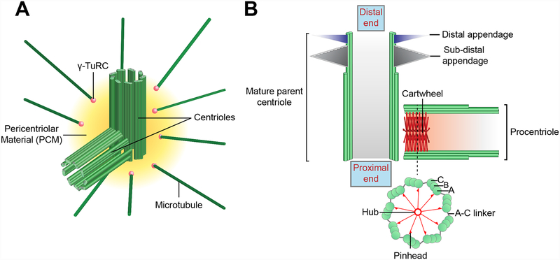
Figure 1. Centriole and centrosome structure.
A) Architecture of the mammalian centrosome. The centrosome is comprised of a pair of orthogonally oriented centrioles surrounded by a proteinaceous Pericentriolar Material (PCM). The PCM contains proteins required for microtubule nucleation and anchoring, such as the γ-Tubulin Ring Complex (γTuRC) (pink spheres). B) Schematic illustration of a mature parent centriole and associated procentriole. Centrioles are cylindrical strictures comprised of nine triplet microtubules, each of which contains a complete A-tubule and an incomplete B and C-tubule. The cartwheel is present in the proximal lumen of the procentriole and is formed by a central hub from which nine spokes emanate. Each spoke terminates in a pinhead structure that binds to the A-tubule of the microtubule triplet. The A-tubule of one triplet is linked to the C-tubule of the adjacent triplet via an A-C linker. Mature parent centrioles are decorated at their distal end with ninefold symmetric distal and sub-distal appendages.

