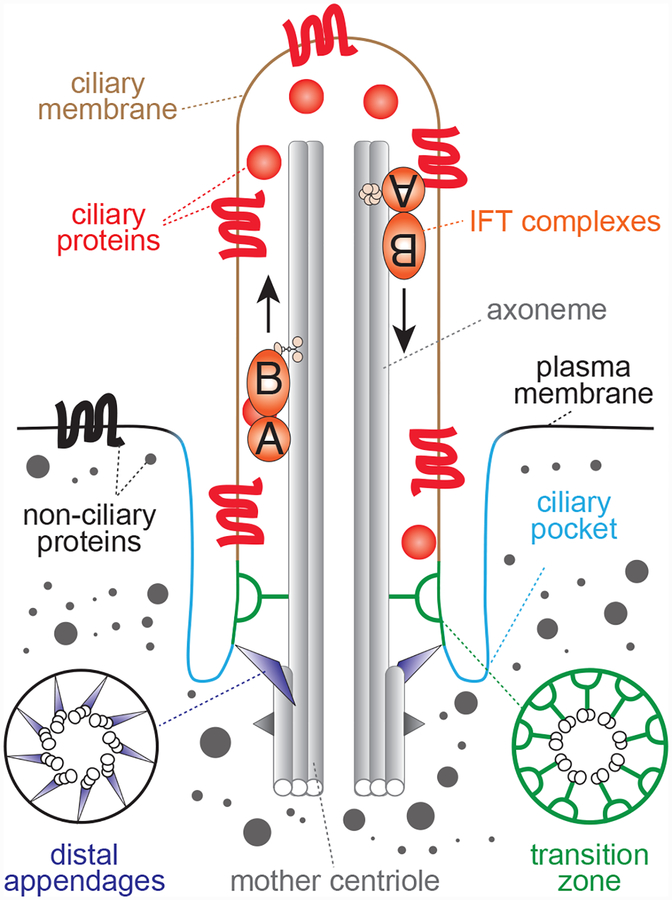
Figure 3. Primary cilium structure.
Architecture of a mammalian primary cilium, highlighting key structural features. The axonemal microtubules form the core of the cilium and extend from the mature parent centriole, which is docked at the plasma membrane. This docking is mediated by the mature centriole’s distal appendages and often occurs at a site on the cell surface where the plasma membrane is invaginated. This invaginated region of the plasma membrane adjacent to the cilium is known as the ciliary pocket is a key site for exo/endocytosis of ciliary materials. Although the cilium lacks a delimiting membrane, it contains a distinct complement of soluble and membrane proteins. This compartmentalization is enabled by diffusion barriers near the base of the cilium at a region known as the transition zone (TZ). The transition zone is made up of several functional and physical modules, including MKS and NPHP proteins, which are mutated in Meckel Syndrome and Nephronophthisis, respectively. Selective trafficking of proteins to cilia across the transition zone is mediated by trafficking machineries, such as IFT-A and IFT-B, that cooperate with ciliary kinesin and dynein motors. Additionally, IFT-A and IFT-B mediate protein transport within cilia along the axonemal microtubules and are required for ciliogenesis.

