FIG. 3.
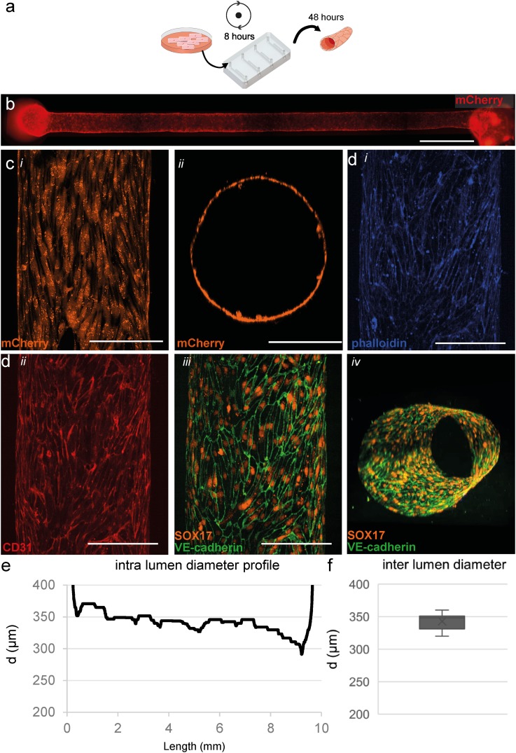
Three-dimensional cell culture of hiPSC-ECs. (a) Schematic overview of cell seeding procedure and culture in microfluidic devices. hiPSC-ECs were seeded and cultured for 48 h in static conditions. (b) Widefield image shows an even and consistent mCherry signal demonstrating uniform coverage of hiPSC-ECs along the whole lumen in a collagen scaffold. (c), (i) Top-down view of live cell confocal image, (ii) XY-reconstruction of the live cell confocal microscopy confirms complete coverage around the perimeter of the lumen. (d) Top-down reconstruction of the lumen visualized using the following markers. (i) F-actin (phalloidin, visualized in blue), (ii) CD31 (visualized in red), and (iii) VE-Cadherin (visualized in green) at the periphery of the hiPSC-ECs costained with SOX17 (visualized in orange) localized at the nuclei of endothelial cells showing alignment with the longitudinal axis of the lumen, and (iv) 3D reconstruction of the engineered vessel showing VE-cadherin and SOX17 around the complete periphery of the lumen and a more detailed reconstruction is presented in video S1. (e) Analyses of the full-length channel show a uniform diameter with small tapering near the outlet. (f) Diameter analysis of cellularised lumens (n = 8), on average 343 ± 12 μm. Scale bars, (b): 1000 μm, (c) and (d): 200 μm.

