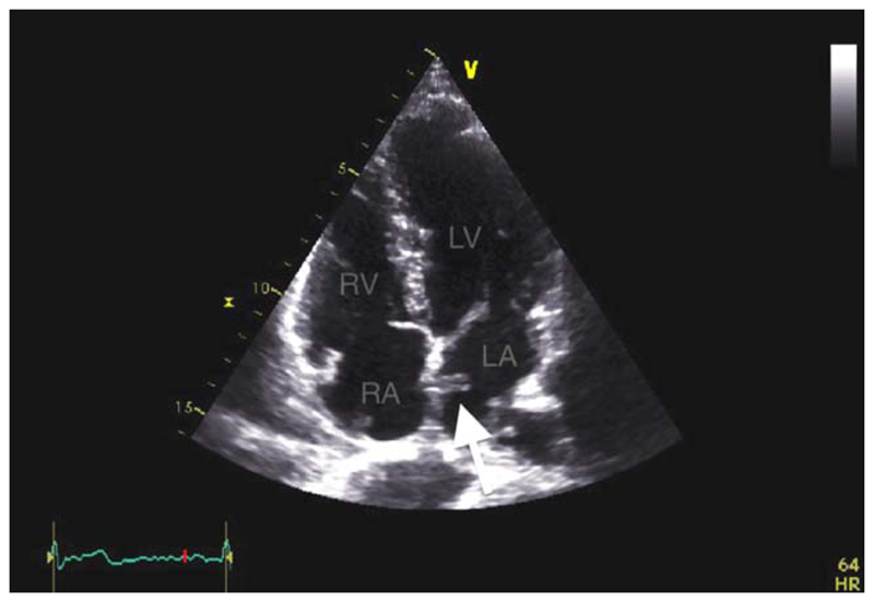
Fig. 7.
Transthoracic echocardiography (TTE). Clot on the left atrial aspect of a GSO occluder (arrowed). LA: left atrium; RA: right atrium; RV: Right ventricule; LV: left ventricle. [Color figure can be viewed in the online issue, which is available at wileyonlinelibrary.com.]

