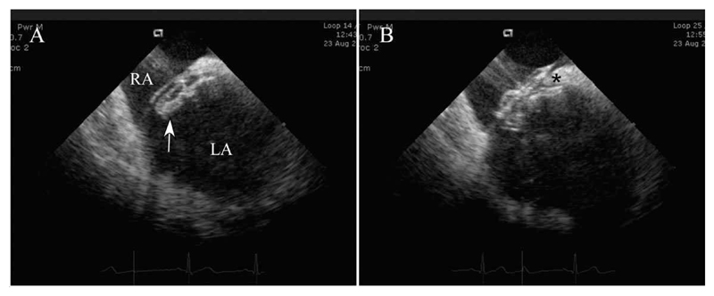
Fig. 2. ICE images of deployment.
A: Left atrial disc (arrow) has been deployed in the left atrium and the whole system has been pulled against the atrial septum. B: The right atrial disc has been deployed, both the septum secundum superiorly (*) and the thinner septum primum inferiorly being held between the two atrial discs of the septal occluder. Abbreviations: LA, left atrium; RA, right atrium.

