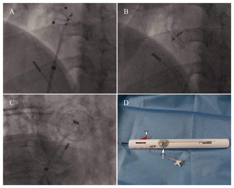
Fig. 3. Fluoroscopy images of deployment.
A and B: LAO projection, C: RAO projection. A: Both left and right atrial discs (*) have been deployed and the occluder lock mechanism has just been pulled back. Three eyelets can be seen from the right to left atrial discs. The intracardiac echo probe tip can be seen in the bottom right corner B: The retention suture has been withdrawn and the delivery catheter has fallen away from the device. C: Image of device in RAO projection showing the radio-opaque petal and circumferential appearance of the nitinol in each disk. D: Appearance of handle assembly post device deployment with the loading and deployment slider at the proximal end of the handle, the retrieval cord lock removed from the slider (arrow) and the suture removed. The occluder locking mechanism slider (arrowhead) has also been pulled back. [Color figure can be viewed in the online issue, which is available at wileyonlinelibrary.com.]

