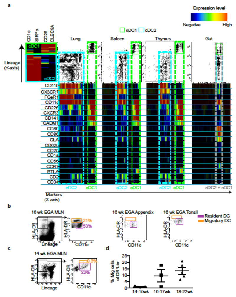
Figure 2. Fetal cDC migrate to draining lymph nodes.
a, Characterisation of cDC1 and cDC2 across fetal tissues using CyTOF and one-Sense algorithm (see Methods, representative plots of n=5). b, c Flow cytometry analysis of fetal mesenteric lymph nodes (MLN) at 16wk (b) or 14wk (c) EGA. Within the HLA-DR+Lin- gate (black), MLN- HLA-DRintCD11chi resident DC (pink) are distinguished from HLA-DRhiCD11int migratory DC (orange gate). b, 16wk EGA MLN (left) and fetal appendix and tonsil (right, n=3). d, Enumeration of migratory cDC at indicated time points. Mean±s.e.m. Each data point in the scatter plots represents an individual experiment.

