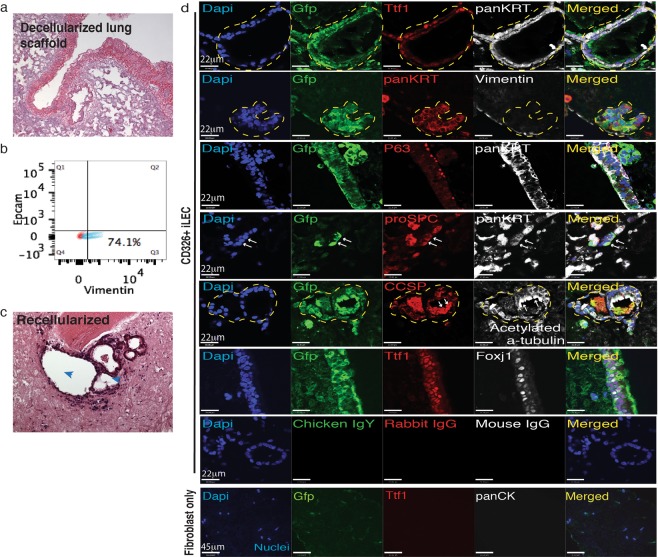
Figure 3.
Lung repopulation potential of mouse iLEC. (a) Hematoxylin and eosin staining of a rat decellularized lung scaffold. (b) Flow cytometric analysis of rat embryonic lung fibroblasts (RELF) for Epcam, epithelial and vimentin, stromal cell markers. (c) Recellularized lungs with iLEC and RELF to provide paracrine support show airway reconstitution and evidence of cilia (blue arrowheads) protruding into the luminal space (right). (d) Immunofluorescence characterization of the recellularized scaffolds show donor mouse iLEC (Gfp+) reconstituting the airway structures with lung (Nkx2-1+) epithelial (panKrt+) cells. Further characterization shows Club (Ccsp+), Ciliated (Acetylated tubulin+, Foxj1+), Basal (P63+), and cells expressing Type II alveolar epithelial cell marker proSPC+ (white arrows). Of note, pan cytokeratin-expressing Gfp+ cells do not express mesenchymal marker Vimentin. No airway epithelial reconstitution was observed when scaffolds were seeded with rat embryonic lung fibroblasts only. Respective non-immune immunoglobulins were used for staining controls. N ≥ 3 recellularization experiments with separate batches of mouse iLEC, RELF and decellularized scaffolds. Scale bars represent 22–45 μm. Yellow hatched indicate GFP+ iLEC areas co-expressing lung markers.

