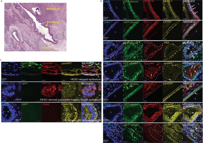
Figure 6.
Human iLEC form lung epithelia in vivo. Human iLEC were co-transplanted subcutaneously into NOD-SCID animals with human embryonic stem cells. (a) Hematoxylin and eosin staining show teratomas containing all three germ layers, ectoderm, mesoderm and endoderm lineages. (b) Immunofluorescence characterization of the teratomas demonstrates presence of human iLEC-derived GFP+ lung epithelial cells expressing NKX2-1 and KRT8/18. Ciliated (FOXJ1+), Basal (P63+), Goblet (MUC5ac+) and Type II alveolar cell (proSPC+) were found lining the iLEC-derived epithelium. Apical expression of CFTR was also detected. White hatched represent GFP+ iLEC areas co-expressing specified markers.White arrows point to positive cells. Yellow arrows point to iLEC-derived (GFP+) mesenchyme (Vimentin+). (c) High magnification of hepatic/pancreatic ductal tissue (KRT19 + Hpd1+, white hatched area) and neuronal (Nestin+) cell clusters were human ES cell-derived. Donor (GFP+) stromal cells (Vimentin+) surround human ES-derived epithelial (panKRT + GFP−). Pink hatched represent GFP− hES-derived areas co-expressing specified markers.Teratoma experiments were replicated n = 2 times with four human iLEC lines. L; lumen. Scale bars represent 22–29 μm.

