Fig. 1.
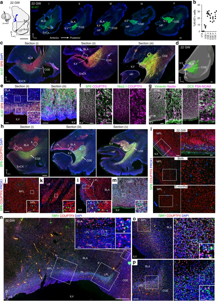
The PL is adjacent to the CGE at 22 gestational weeks (GW). a (Left) 22 GW human brain, coronal section of the basolateral amygdala (BLA). (Right) Ki-67+ cells in temporal lobe sections (i–v) spanning the anterior BLA to the caudal ganglionic eminence (CGE), including the entorhinal cortex (EnCX), and hippocampus (HP). b Percentage of Ki-67+ DAPI-stained nuclei in the temporal lobe cortex (CTX) or in the CGE in each section from a. (Left to right, n = 4, 11, 10, 4, 5, and 4 independent z-stacks analyzed per section, error bars s.e.m.). c Ki-67+SP8+ cells beneath the amygdala and surrounding the anterior tip of the temporal horn of the lateral ventricle (tLV) in sections i–iii from a. d 3-D reconstruction of sections in a showing the CGE extending beneath the BLA and the PL. e Boxed regions in sections ii and iii from c showing (Left) few Ki-67+SP8+ cells in the PL, more between the PL and ventricle, and (Right) many in the CGE. f SP8+COUP-TFII+ cells and few NKX2.1+ cells in the CGE at 22 GW. g Vimentin+nestin+ and DCX+PSA-NCAM+ cells in the CGE at 22 GW. h (Top) Coronal sections adjacent to i, iii, and v from a showing COUP-TFII+SP8– cells in the BLA, and COUP-TFII+SP8+ cells in the CGE. i COUP-TFII+SP8–PROX1– cells in the MPL at 22 GW, birth, and 5 months postnatal. j–m Higher magnification of boxed regions in h. COUP-TFII+SP8–PROX1– cells in the (j) medial PL (MPL), (k) lateral amygdala (LA), and (l) lateral PL (LPL) next to (m) COUP-TFII+SP8+PROX1+ cells in the CGE. n–p COUP-TFII+ cells in the (o) MPL and (p) LPL express TBR1 mRNA, unlike the COUP-TFII+ cells in the adjacent CGE (inset). Scale bars: 10 mm (a left), 1 mm (a right, c, d, h), 500 µm (n), 100 µm (e, f left, i left, j–m, o left, p left), 20 µm (e inset, f right, g right, i right, j–m insets, n left inset, o right, p right), 10 µm (n–p right insets)

