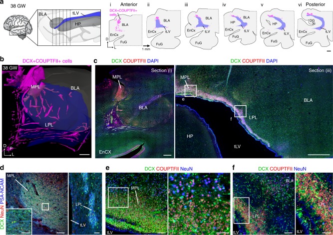
Fig. 2.
Distribution of DCX+COUP-TFII+ cells in the PL at birth. a Schematic of 38 GW temporal lobe indicating approximate level of coronal sections separated by 1 mm. Maps of the locations of dense clusters of DCX+COUP-TFII+ cells on the medial and ventral sides of the amygdala beginning at the anterior tip of the temporal lateral ventricle (tLV) corresponding to coronal section (i). b 3-D reconstruction of DCX+COUP-TFII+ clusters (purple) around and within the BLA (gray). c DCX+COUP-TFII+ cells in the PL at anterior and posterior levels (i and iii). d DCX+PSA-NCAM+ cell clusters are mostly NeuN– in the MPL and LPL at birth. e, f DCX+COUP-TFII+NeuN– cells in the MPL and LPL at birth from sections adjacent to the boxed areas in (c, iii). Fusiform gyrus (FuG). Scale bars: 2 mm (a), 1 mm (b, c), 200 µm (d left), 100 µm (e left, f left), 50 µm (d left inset, d right, e right, f right)

