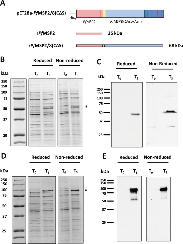
Figure 1.
Expression of unfused rPfMSP2 and chimeric rPfMSP2/8. (A) Cartoon of expression constructs for rPfMSP2 and rPfMSP2/8 production. Cysteine residues are indicated by red bars, yellow region indicates Gly-Ser linker domain. (B,C) E. coli lysate at time of induction (T0) or 3 hours post-induction (T3) was separated on 10% polyacrylamide gels and rPfMSP2 expression was analyzed by (B) Coomassie Blue staining and (C) immunoblot using anti-PfMSP2 antibody (1:20,000 dilution). (D,E) rPfMSP2/8 expression was analyzed by (D) Coomassie Blue staining and (E) immunoblot as described for rPfMSP2. Asterisks indicate induced rPfMSP2 and rPfMSP2/8 protein products. The T0 samples served as negative controls for the immunoblot analysis of protein expression.

