Figure 3.

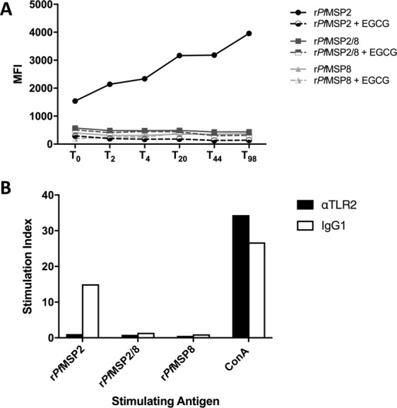
Assessment of fibril formation and stimulation of splenocytes with rPfMSP2 antigens. (A) Recombinant proteins (10 μM) were stained with ThT (30 μM) and incubated at 37 °C. Fluorescence was measured at baseline (T0) prior to incubation and at indicated time points throughout (T2-T98, solid lines). In parallel, EGCG (40 μM) was co-incubated with recombinant proteins to prevent fibril formation and measured as above (dashed lines). Data are presented as the average of duplicate samples. (B) Naïve mouse splenocytes were plated, and triplicate samples were stimulated with recombinant antigen (10 μg/ml) in the presence of 1 μg/ml neutralizing anti-TLR2 mAb (black bars) or an IgG1 isoptype control (white bars). Concanavalin A (ConA, 1 μg/ml) served as positive control. Proliferation was quantitated by [3H]thymidine incorporation, and stimulation index was calculated for each sample relative to unstimulated controls.
