Fig. 5.
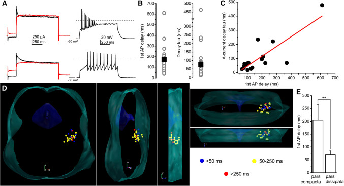
Neurons possessing transient outward current can be grouped as early- and late-firing neurons. a Current-and voltage traces recorded from an early-(upper row) and a late-firing neuron (lower row). Left: current traces elicited with depolarizing square voltage steps, preceded by hyperpolarizing (black) and depolarizing prepulses (red). Right: voltage traces recorded with 100 pA depolarizing square current injections from − 80 mV resting membrane potentials. b Statistical summary of the spike latency (recorded at − 80 mV resting membrane potential) and the decay tau of the declining phase of the transient outward currents (gray circles: individual data; black squares: average ± SEM). c Decay tau of the transient outward currents plotted against the spike latency at − 80 mV resting membrane potential (individual data: black squares; red line: linear fit). d Distribution map of spike latencies at − 80 mV resting membrane potentials (blue: latency below 50 ms—considered as neurons with no transient outward currents, yellow: latency between 50 and 250 ms—considered as early-firing neurons; red: longer latency than 250 ms—considered as late-firing neurons). Note that late-firing neurons are located caudally. e Statistical comparison of spike latencies from the pars compacta (caudal region) and pars dissipata (rostral region; average ± SEM)

