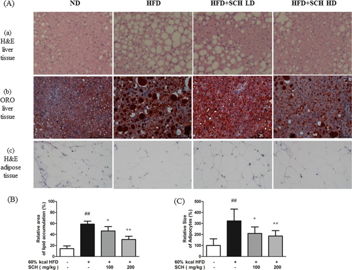
Figure 9.
Histologic analysis of tissues showing effects of 15 week SCH administration on hepatic lipid accumulation and size of epididymal adipocytes of HFD-fed c57BL/6 mice. (A) Representative images of H&E stained liver tissue (a), Oil Red O stained liver tissue (b), and H&E stained adipose tissue (c) from each group. Quantification of lipid stained area of liver tissue (B) and average size of adipocytes (C). Data are expressed as mean ± SD.

