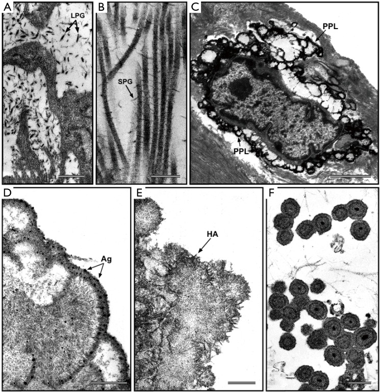
Figure 2.
Ultrastructural visualization of polyanionic compounds after pre-embedding histochemical reaction with phthalocyanin cuprolinic blue (CB). (A) CB-positive interstitial leaf-like large proteoglycans (LPG) and (B) CB-positive rod-like small proteoglycans (SPG) interconnecting adjacent collagen fibrils, in native aortic valve cusps. (C) Phthalocyanin-positive layer (PPL) edging a calcifying interstitial cell in an aortic valve cusp subjected to in vivo experimental calcification. (D) Peripheral PPL showing superimposed silver particles (Ag) after additional post-embedding von Kossa reaction in a degenerating aortic valve interstitial cell (AVIC) subjected to in vitro calcification. (E) Peripheral precipitation of needle-like hydroxyapatite (HA) crystals at the level of underlying PPL in a degenerating AVIC subjected to in vitro calcification. (F) PPL-lined calcospherulae originated from vesicular remnants released by AVICs subjected to in vitro calcification. Bar: 0.5 µm (A); 0.25 µm (B); 25 µm (C); 0.5 µm (D); 0.5 µm (E); 1 µm (F).

