
Figure 2.
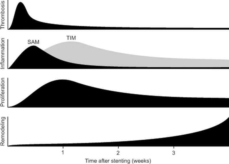
Activity of the four primary components of arterial injury following stent placement. Platelet-rich thrombosis peaks 3–4 days after stent deployment especially over areas of strut injury. Concomitant inflammation is initially mediated by surface-adherent monocytes (SAM) recruited to the injury site that then migrate into the neointima as tissue-infiltrating monocytes (TIM) and accumulate around the stent struts as giant cells. Vascular smooth muscle cell proliferation peaks 7 days after stent deployment coincident with the transition of SAM to TIM and continues for weeks afterward. Extracellular matrix deposition in the adventitia, tunica media, and neointima accelerates at week 3 after stent deployment and underlies arterial remodeling and subsequent luminal narrowing. Adapted from Edelman and Rogers.36