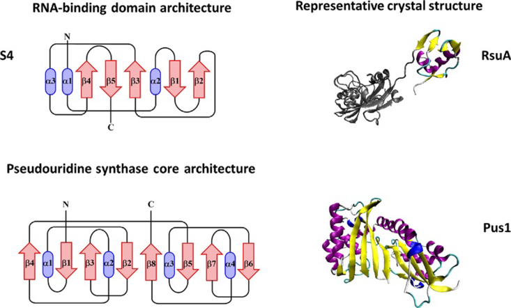
Fig. 5.
Topological diagram of the S4 RNA-binding domain (top left) and representative crystal structure of RsuA containing an S4 domain (top right). The protein structure outside of the featured domain is shown in gray cartoon. Topological diagram of the core domain of pseudouridine synthases based off of the E. coli enzyme TruA (bottom left). The core of pseudouridine synthases is comprised of two domains each with a βαββαβ arrangement forming an alternating eight-stranded β-sheet. The arrangement, but not necessarily the connectivity shown here, is conserved among pseudouridine synthases. Representative crystal structure of human Pus1 (bottom right). These models were created using the Visual Molecular Dynamics (VMD) program [52].

