
Fig. 16.
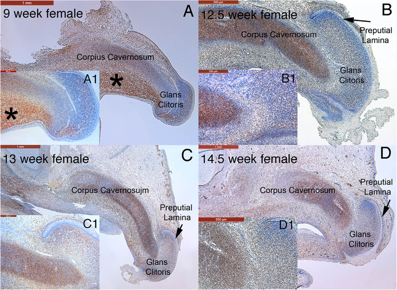
Mid-sagittal sections stained for the androgen receptor in the developing human female genital tubercle at 9 weeks of gestation (A) and clitoris at 12.5, 13 and 14.5 weeks of gestation (B-D, respectively). Note the strong localization of the androgen receptor to the corpus cavernosum and glans clitoris at all fetal ages. In the 9-week specimen note especially strong staining of the androgen receptor in the mesenchyme below the corpus cavernosum (* in A). A1-D1 are high power images of each time point.