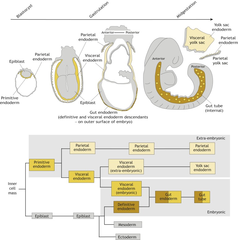
Fig. 2.
The endoderm of the mouse embryo arises from two sources with distinct developmental origins. Schematic overview (top) and lineage tree (bottom) depicting the development of endodermal tissues in mice, from the blastocyst stage to the midgestation embryo. The gut endoderm forms on the surface of the embryo at gastrulation where definitive endoderm (derived from the epiblast; brown) cells intercalate (egress; Schöck and Perrimon, 2002) into the overlying visceral endoderm (derived from the primitive endoderm, yellow). The gut endoderm then becomes internalized and forms the gut tube and will give to the epithelial lining of all endodermal tissues in the adult organism. The gut tube therefore comprises cells of two different origins: extra-embryonic (beige) and embryonic (yellow). Parietal and yolk sac endoderm (beige), which are also derived from primitive endoderm in the blastocyst, solely give rise to extra-embryonic structures.

