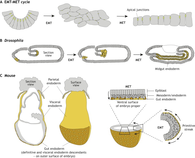
Fig. 3.
The EMT-migration/translocation/relocation-MET cycle in Drosophila and mouse embryos. (A) Schematic of cells undergoing an EMT-MET cycle. Cells within an epithelium undergoing EMT lose their apico-basal polarity and loosen their cell-cell junctions (green). After undergoing EMT, cells display a mesenchymal morphology and adopt migratory behaviour. As they rejoin an epithelium and undergo MET, they re-establish cell polarity (by upregulating apico-basal polarized proteins) and reform or reinforce cell-cell junctions. (B) In Drosophila, an EMT-MET cycle occurs during midgut formation. Future endoderm cells delaminate from the anterior and posterior poles of the embryo. Cells from both sections migrate and meet to form the midgut section of the gut. Through interactions with the underlying mesoderm, endoderm cells undergo MET and repolarize to form the midgut epithelium. (C) In mice, an EMT-MET cycle occurs during gastrulation. Definitive endoderm cells (brown) undergo EMT when they leave the primitive streak and migrate along the wings of mesoderm. They then undergo MET as they intercalate in the overlying visceral endoderm (yellow) epithelium to form the gut endoderm. Section and surface whole-mount views show the location of the gut endoderm on the surface of the mouse embryo, consisting of definitive endoderm cells and visceral endoderm cells. Cross-sections through the embryo show the position of an EMT at the primitive streak, and an MET during gut endoderm formation.

