Fig. 5.

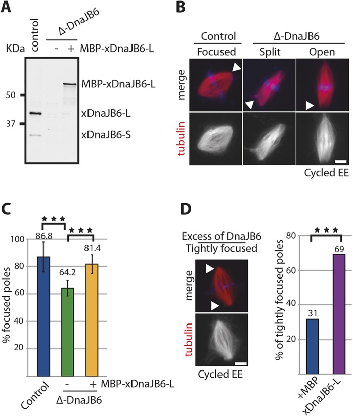
DnaJB6 depletion from Xenopus egg extracts induces spindle pole organization defects. (A) Western blot analysis of control and xDnaJB6-depleted (Δ-DnaJB6) Xenopus egg extracts (EE) without (−) or with (+) addition of the recombinant MBP–xDnaJB6-L. The depletion was very efficient and the recombinant protein was added at close to endogenous concentrations. 1 μl of egg extract was loaded per lane. The endogenous and recombinant proteins were detected with an in-house generated anti-xDnaJB6 antibody. (B) Fluorescence images of spindles assembled in control or DnaJB6-depleted egg extracts. Most spindles assembled in control-depleted extracts have focused spindle poles, whereas many spindles assembled in DnaJB6-depleted extracts show spindle pole focusing defects defined as ‘Split poles’ or ‘Open poles’ as shown. The white arrowheads point to the different types of spindle poles. In the merge, tubulin is shown in red and DNA in blue. Scale bar: 10 μm. (C) Graph showing the mean±s.d. percentage of focused spindle poles in control extracts (blue), DnaJB6-depleted extracts (green) and DnaJB6-depleted extracts containing MBP–xDnaJB6 (orange). Data are from four independent experiments in which 703, 607 and 358 spindle poles were examined, respectively. ***P<0.001 (ANOVA test). (D) Fluorescence image of a spindle assembled in egg extract supplemented with an excess of DnaJB6 (1 μM) showing tightly focused spindle poles (white arrowheads). Scale bar: 10 μm. Bar graph on the right shows the percentage of tightly focused poles in spindles assembled in control extracts supplemented with MBP (blue) and in extracts supplemented with MBP–xDnaJB6 (purple). Data are from three independent experiments in which 114 and 258 spindle poles were analyzed. ***P<0.0001 (Fisher exact test).
