Fig. 6.
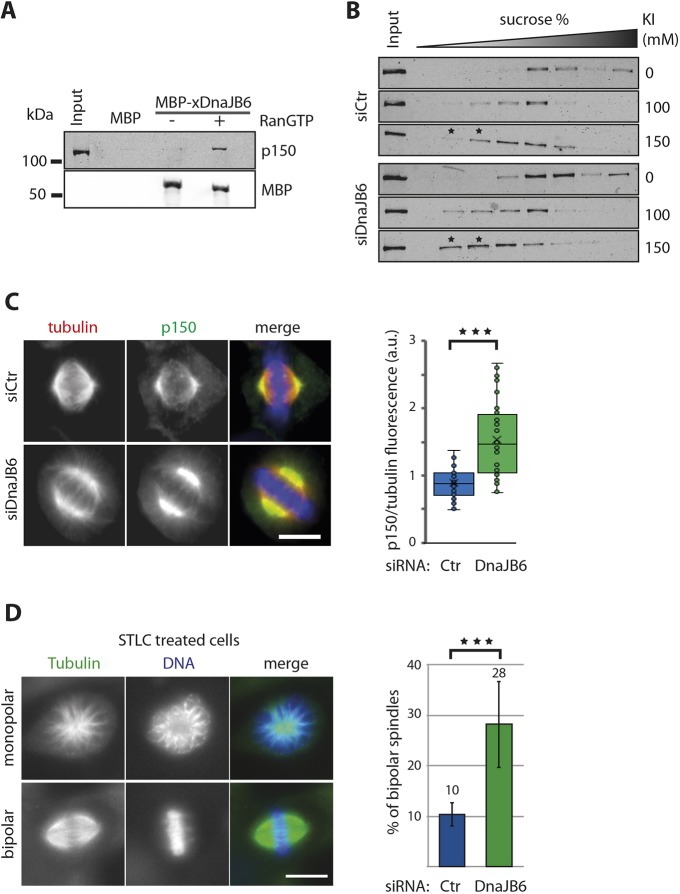
DnaJB6 regulates dynactin spindle localization and is required for dynein-dependent force generation within the spindle. (A) Western blot analysis of a MBP–xDnaJB6-L pulldown experiment in Xenopus egg extract. MBP–xDnaJB6-L- or MBP-coated Dynabeads were incubated in egg extracts in the presence or absence of RanGTP. p150Glued was specifically pulled down with MBP–xDnaJB6-L from extracts containing RanGTP. The lower panel shows that similar amounts of MBP–xDnaJB6-L was used for pulldown both in the presence and absence of RanGTP. (B) Western blot analysis showing the position of p150Glued in 8–20% sucrose density gradients from lysates of control and DnaJB6-silenced cells. Lysates were prepared from mitotic cells. KI was added to the lysates and incubated for 1 h before running the gradients, at the concentrations indicated on the right. A shift to in where p150Glued is detected in to fractions with a lower percentage of sucrose is observed in mitotic DnaJB6 cell lysates treated with 150 mM KI compared to the mitotic control cells lysates with the same treatment, as highlighted with asterisks. (C) p150Glued accumulates at spindle poles in DnaJB6-silenced cells. Left: representative immunofluorescence images from control and DnaJB6-silenced HeLa cells showing the localization of p150Glued in metaphase spindles. In the merge, tubulin is in red, dynein is in green and DNA is in blue. Scale bar: 10 μm. Right: box-and-whisker plot showing the value of the of p150Glued signal normalized to that of the tubulin signal (a.u., arbitrary units) in each spindle pole in control and DnaJB6-silenced HeLa cells. More than 30 cells were analyzed for each condition in one representative out of three independent experiments. (D) DnaJB6 silencing rescues spindle bipolarity in STLC-treated HeLa cells. Left: immunofluorescence images of representative monopolar and bipolar spindles in DnaJB6-silenced HeLa cells incubated with STLC. Tubulin is shown in green and DNA in blue. Scale bar: 10 μm. Right: bars graph showing the mean±s.d. percentage of bipolar spindles in control or DnaJB6-silenced HeLa cells incubated with STLC. Data from three independent experiments in which 937 control and 1057 DnaJB6-silenced cells were analyzed. ***P<0.001 (ANOVA test).

