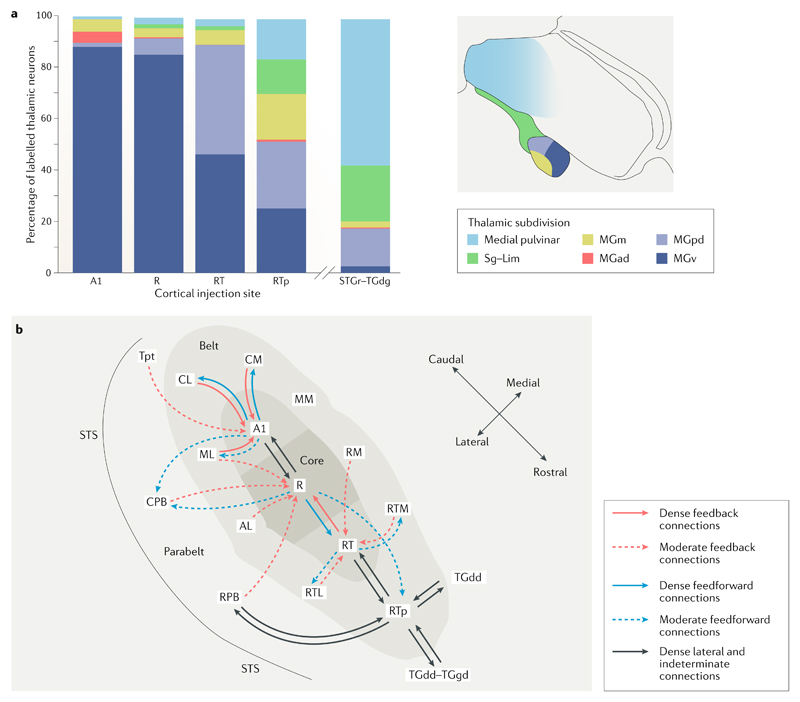
Fig. 1. Cortical and subcortical connectivity of the macaque auditory cortex.
a | Distribution of inputs to each auditory cortical field from the different thalamic nuclei. There is a clear rostral–caudal shift in thalamic connectivity. Moving rostrally, there is a general decline in the proportions of connections from the ventral division of the medial geniculate nucleus (MGN) of the thalamus (MGv) and increased proportions of inputs from other MGN and other thalamic nuclei. Core areas A1 and R receive an overwhelming majority of their inputs from the MGv, whereas the more rostral temporal core field (RT) area receives similar proportions of inputs from the MGv and the posterodorsal MGN (MGpd). The rostrotemporal polar belt area (RTp) receives roughly similar proportions of its inputs from the MGv, MGpd and the medial division of the MGN (MGm) as well as the suprageniculate (Sg)–limitans (Lim) complex and the medial pulvinar. The rostral superior temporal gyrus (STGr) and the granular part of the dorsal temporal pole (TGdg) receive the majority of their thalamic inputs from the medial pulvinar and a lower proportion from the Sg–Lim complex and the MGpd13. b | A schematic image illustrating the connectivity of different core auditory regions in the macaque cortex12,19,111. Dense connections (those for which the retrograde connectivity cell count was over 30) are represented with solid lines, whereas moderate connections (those for which cell count was between 15 and 29) are shown with dashed lines. The connectivity pattern shows a clear rostral–caudal difference. Caudal core field A1 primarily connects to surrounding belt fields and to the rostral auditory core field (R), with more moderate connections to the caudal belt and parabelt fields. R, on the other hand, connects to A1 and to the RT, with moderate connections to the rostral and caudal belt and parabelt fields and the RTp. RT connects to the adjacent field RTp and to the adjacent rostral belt fields. RTp has a distinctly different pattern of connectivity from the temporal pole, rostral belt and parabelt fields via lateral and indeterminate connections. This pattern of connectivity results in a recurrent and interactive network incorporating multiple parallel pathways with both direct and indirect connections12. AL, anterolateral belt; CL, caudolateral belt; CM, caudomedial belt; CPB, caudal parabelt; MGad, anterodorsal division of the MGN; ML, middle lateral belt; MM, middle medial belt; RM, rostromedial belt; RTL, rostrotemporal-lateral belt; RTM, rostrotemporal-medial belt; RPB, rostral parabelt; STS, superior temporal sulcus; TGdd, dysgranular part of the dorsal temporal pole; Tpt, temporo-parietal area. Part a is adapted with permission from REF.13, Wiley-VCH. Part b is adapted with permission from Scott, B. H. et al. Intrinsic connections of the core auditory cortical regions and rostral supratemporal plane in the macaque monkey. Cereb. Cortex (2017) 27(1): 809–840, by permission of Oxford University Press (REF.12).

