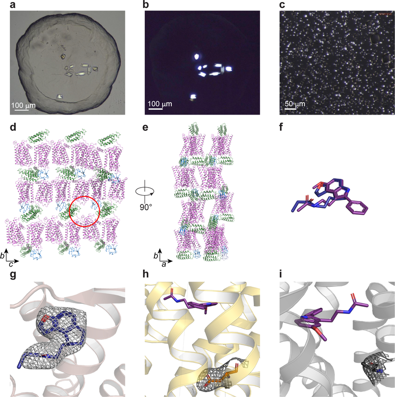
Extended Data Fig. 1 |. Crystallisation of MT2: crystals, crystal packing, and electron density.
a, Bright field and b, cross-polarised images of representative MT2-2-pmt crystals optimized for synchrotron data collection (representing three independent crystallisation setups). c, cross-polarised image of representative MT2-N86D-2-pmt crystals used for XFEL data collection (representing three independent crystallisation setups). See Extended Data Table 2 for data collection statistics. d, e, Crystal packing (receptor - purple, BRIL – green, and rubredoxin - blue). Space for missing rubredoxin in molecule B of the asymmetric unit is indicated with a red circle. Lattice rotated 90° is shown in e. f, Overlay of 2-pmt (purple) and ramelteon (blue) ligands of MT2. g-e, 2mFo-DFc density (grey) contoured at 1 σ of ramelteon (g), N862.50D mutation (h), and H2085.46A mutation (i). 2-pmt is shown in purple.

