
Fig. 1.
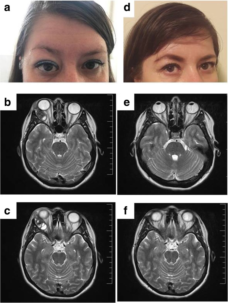
a Preoperative photograph of the patient showing right exophthalmia. b, c MRI detected a well-defined 4 × 2,2 × 2,7 cm mass in the right lacrimal gland region that showed a septate pseudocystic appearance and exerted a mass effect on adjacent structures without local bony destruction. d Postoperative case photograph showing normalization of the globe and resolution of right exophthalmia. e, f No evidence of residual tumour was found in the orbital MRI control