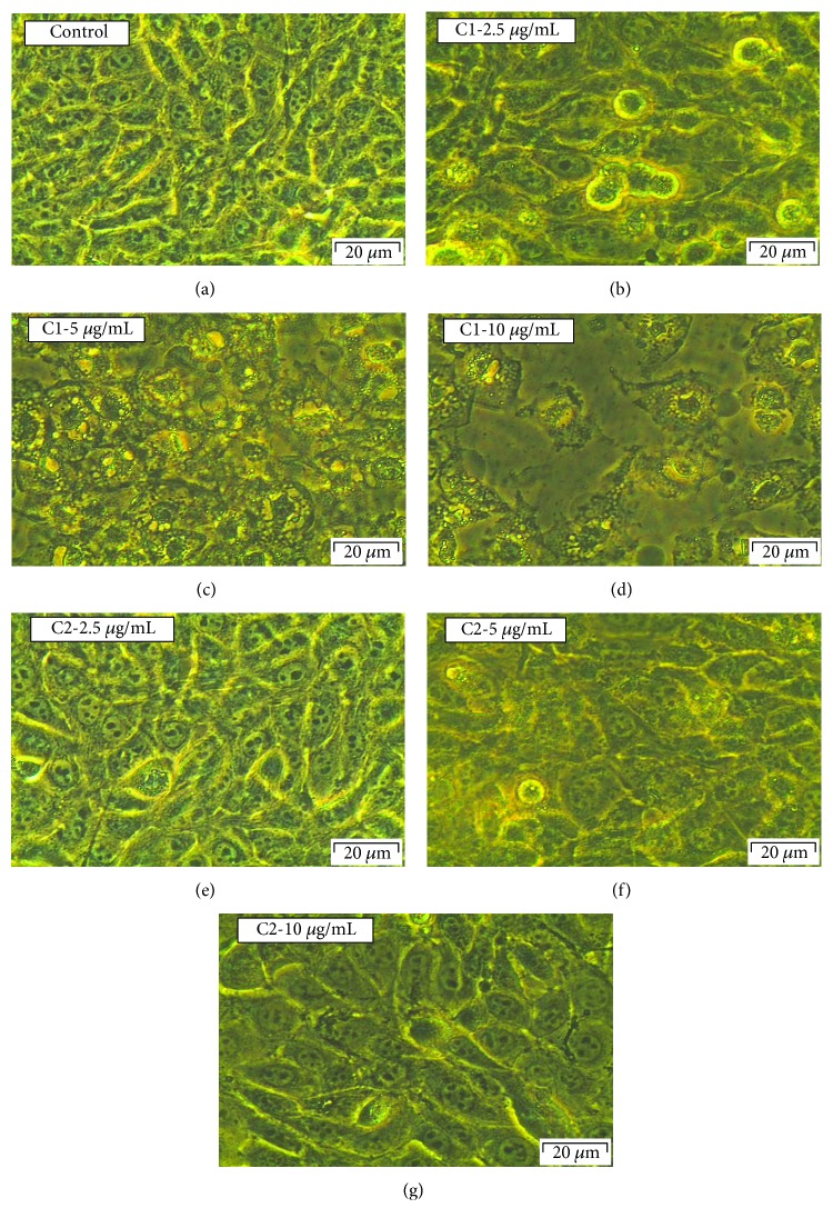
Figure 2.
Morphological evaluation of C1- and C2-treated MCF-7 cells. There were no significant visible differences in control (a) and 2.5 μg/mL C2-treated groups (e); the cells did not show any cellular shrinkage and apoptotic bodies after the treatment. However, more dead cells were observed at all concentrations of C1 and higher concentration of C2 (5 and 10 μg/mL). The treated cells showed loss of intact membrane and loss of contact with neighbouring cells and were condensed; being detached from the culture plate showed the features of apoptotic cells.

