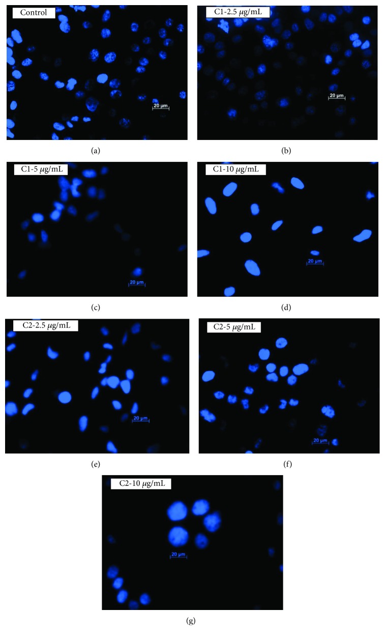
Figure 8.
Hoechst staining. Nuclear damage was analysed by Hoechst stain. Control cells (a) showed intact nucleus without any damage; however, the cells treated with C1 and C2 (b–g) at different concentrations showed highly condensed chromatin granules, loss of nuclear shape which indicates the DNA damage. C1-treated cells showed higher nuclear damage.

