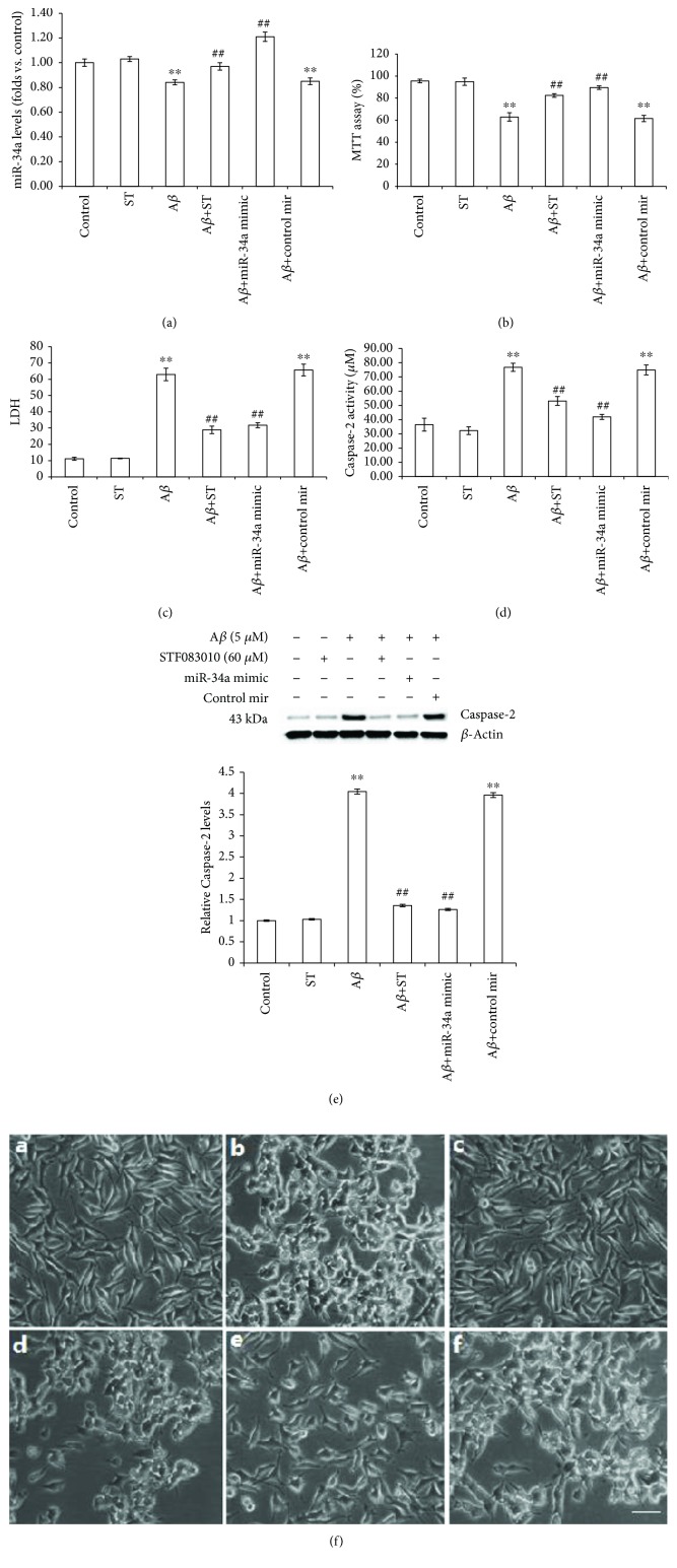
Figure 4.
The roles of IRE1α and miR-34a in Aβ-induced toxicity in SH-SY5Y cells. Cells were incubated with STF-083010 (60 μM)+Aβ1-40 (5 μM), STF-083010 (60 μM) alone, or Aβ1-40 (5 μM) alone or were untreated. In transfection studies, cells were transfected with the miR-34a mimic oligonucleotide or nonspecific control in the presence or absence of Aβ1-40 (5 μM). (a) miR-34a levels were measured using quantitative real-time PCR. (b) Cell viability was measured using the MTT assay. (c) The integrity of the plasma membrane was assessed by LDH release. (d) Caspase-2 activity was measured using the Caspase-2 cellular activity assay kit. (e) The expression of Caspase-2 was measured by Western blotting. (f) Morphological analysis of SH-SY5Y cells by microscopy. Scale bar: 100 mm. Data are presented as the means ± SEM from four independent experiments. ∗∗P < 0.01 compared with the control; ##P < 0.01 compared with the Aβ1-40-alone group.

