Figure 1.
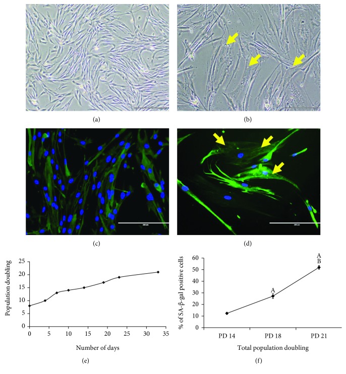
Morphological changes and serial passaging of myoblast cells in culture. Myoblast cells exhibited different morphological characteristics, as seen in the photomicrographs of young (a) and senescent (b) myoblast cells (magnification: 50x), and the photomicrographs of desmin staining of young (c) and senescent (d) cells (magnification: 200x). Myoblasts were stained for desmin (green) and nuclei (blue). Arrows indicate the intermediate filaments and vacuoles observed in senescent myoblast cells. Myoblast cells also lost their proliferative capacity with serial passaging as observed in the proliferation-lifespan curve of myoblast cells (e) and in the increased percentage of cells that stained positive for SA-β-gal at higher PDs (f). The data are presented as the means ± SD, n = 3. Ap < 0.05: significantly different compared to myoblasts at PD 14 (young); Bp < 0.05: significantly different compared to myoblasts at PD 18 (presenescent), with a post hoc Tukey HSD test.

