Figure 5.
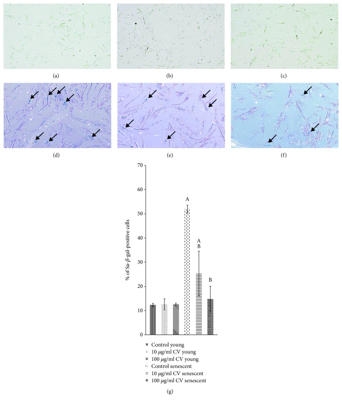
C. vulgaris treatment decreased the percentage of SA-β-gal-positive senescent cells. SA-β-gal staining was used as a senescence biomarker, shown in photomicrographs of young control cells (a), young cells treated with 10 μg/ml C. vulgaris (b), young cells treated with 100 μg/ml C. vulgaris (c), senescent control cells (d), senescent cells treated with 10 μg/ml C. vulgaris (e), and senescent cells treated with 100 μg/ml C. vulgaris (f) (magnification: 40x). This data was quantified by the percentage of cells that stained positive for SA-β-gal (g). The data are presented as the means ± SD, n = 3. Ap < 0.05: significantly different compared to young controls; Bp < 0.05: significantly different compared to senescent controls, with a post hoc Tukey HSD test.

