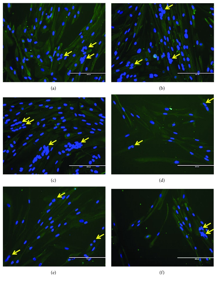
Figure 6.
Desmin staining of differentiated myoblasts, indicating the presence of multinucleated cells in mature myotubes. Photomicrographs of desmin staining on day 3 for young control myoblasts (a), young myoblasts treated with 10 μg/ml C. vulgaris (b), young myoblasts treated with 100 μg/ml C. vulgaris (c), senescent control myoblasts (d), senescent myoblasts treated with 10 μg/ml C. vulgaris (e), and senescent myoblasts treated with 100 μg/ml C. vulgaris (f) (magnification: 200x). Myoblasts were stained for desmin (green) and nuclei (blue). Arrows indicate the multinucleated cells formed during the differentiation and fusion process.

