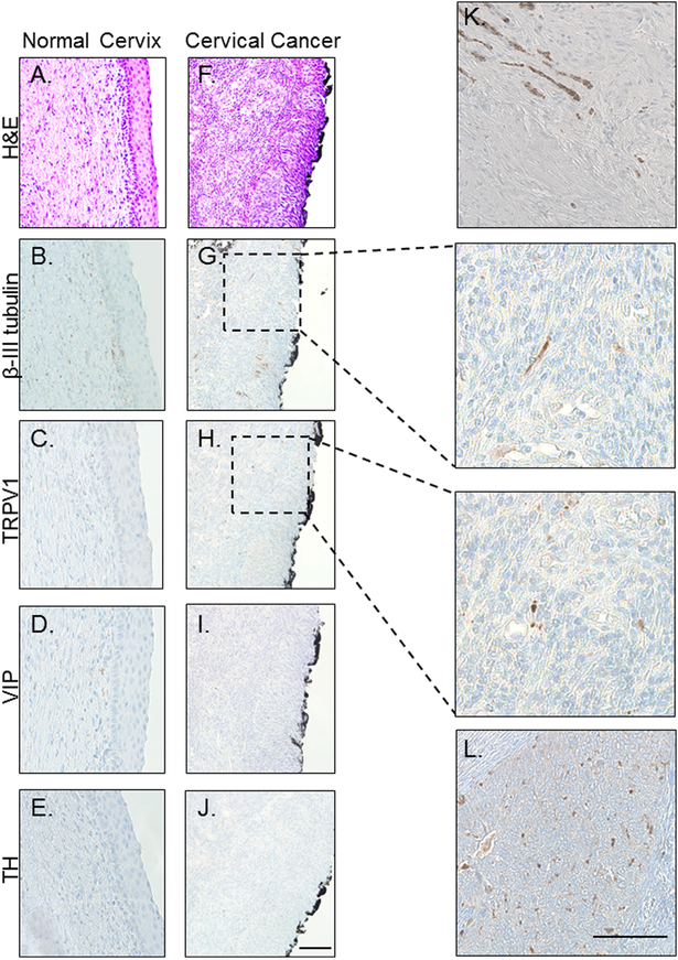
Figure 1: Cervical cancer is innervated.
Representative bright field images of normal cervix (A-E) and cervical cancer (F-L) stained with H&E (A,F) and immunohistochemically stained for β-III tubulin (B,G), TRPV1 (C,H), VIP (D,I) and TH (E,J.). Panels K and L are examples of robust β-III tubulin and TRPV1 staining, respectively. Scale bars, 50 μm. Black boarding the cervical cancer is ink from the surgical procedure.

