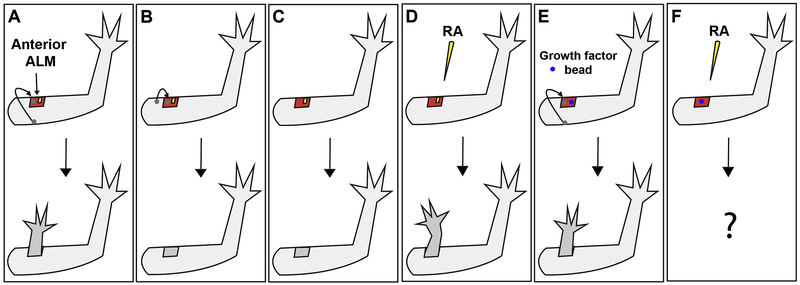
Figure 1: Growth factor replacement of tissue-specific signaling during limb regeneration.
(A) Ectopic limb structures are generated from an anterior-located wound site with a nerve (yellow) and a graft of full-thickness skin (grey circle) from the posterior side of the limb (Endo et al., 2004). (B) A regenerative response is not induced by the same manipulation as in “A” when the grafted tissue is obtained from the anterior side of the limb. (Phan et al., 2015) (C) A wound site with only a nerve (no graft) does not result in regeneration (Endo et al., 2004). (D) Treatment of a wound site with only a nerve, with retinoic acid (RA) results in the formation of ectopic limbs. (McCusker et al., 2014) (E) Implantation of a gelatin bead soaked in FGF-2, FGF-8, and BMP-2 into an anterior-located wound with a tissue graft (no nerve) results in the formation of limb structures (Makanae et al., 2014). (F) In the current study, we are combining the treatments described in “D” and “E” to determine whether ectopic limbs can be induced in superficial wounds treated consecutively with growth factors.

