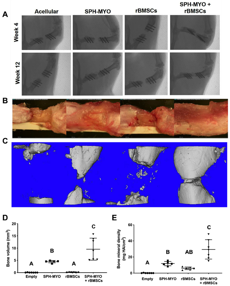
Figure 5. Repair of rat femoral defect when treated with conditioned media.

(A) Week 4 and 12 x-rays of the defect. (B) Images of explanted femurs at week 12. (C) MicroCT images of bone mineralization at week 12 and quantitative microCT data for (D) bone volume and (E) bone mineral density. Bars that do not share letters are statistically significant from each other (n=5-7).
