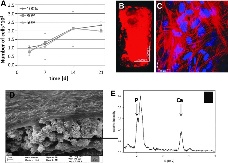
FIG. 18.
Ovine MSCs were cultured on LCM_8:2 scaffolds of different stiffness for 7 days in normal cell culture medium and for additional 14 days in differentiation medium. (A) Time-dependent proliferation of oMSCs in LCM_8:2 100%, LCM_8:2 80%, and LCM_8:2 50% scaffolds; (B) confocal fluorescence image of a whole LCM_8:2 scaffold; and (C) magnification in the interior of unit cell of LCM_8:2 scaffold after culture time of 21 days (red: actin, blue: nucleus); (D) SEM image of LCM_8:2 scaffold cultured with MC3T3–E1 osteoblasts for 21 days; (E) EDX spectrum of calcified area inside the scaffold. EDX, energy dispersive X-ray spectroscopy.

