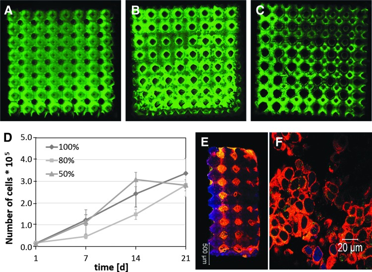
FIG. 20.
Confocal fluorescence microscopy images of (A) LCM_2:8 100%, (B) LCM_2:8 80%, and (C) LCM_2:8 50% scaffolds written by 2-PP with 8 × 8 × 3 Schwarz P unit cells (dimension: 520 μm; overlap 20 μm); MCF-7 cells were cultured on LCM_2:8 scaffolds of different stiffness for 21 days. (D) Proliferation kinetic of MCF-7 cells in LCM_2:8 100%, LCM_2:8 80%, and LCM_2:8 50% scaffolds; (E) confocal fluorescence image of a whole LCM_2:8 scaffold and (F) magnification inside one pore of LCM_2:8 scaffold after culture time of 21 days (red: actin, green: CD44, blue: nucleus). The percentage value indicates the amount of bifunctional copolymer.

