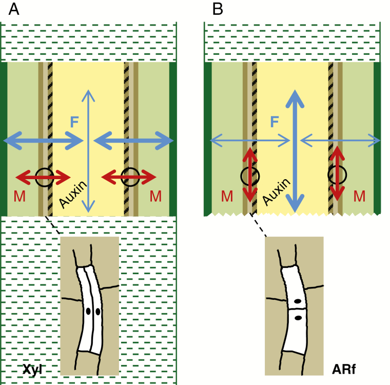
Fig. 1.
Conceptual model of mechanic effects on microtubule orientation and the resulting orientation of cell division in cuttings. In the stem bases of shoot tips, starting from the outside, the different colours represent the epidermis, cortex, phloem, cambium, xylem and pith tissues. Dashed zones illustrate the apical (A, B) and basal (A) stem connected to the stem bases when the shoot tips are attached to the stock plant (A) and after excision (B). Black circles indicate exemplary positions of cambium cells, while the sketches shown below illustrate their periclinal (A) vs. anticlinal (B) cell division. Blue arrows indicate the direction of mechanical gradients, while the thickness of lines indicates the magnitude. Red arrows indicate the orientation of microtubules in cambium cells. F, mechanical forces; M, microtubules; Xyl, xylogenesis; ARf, AR formation.

