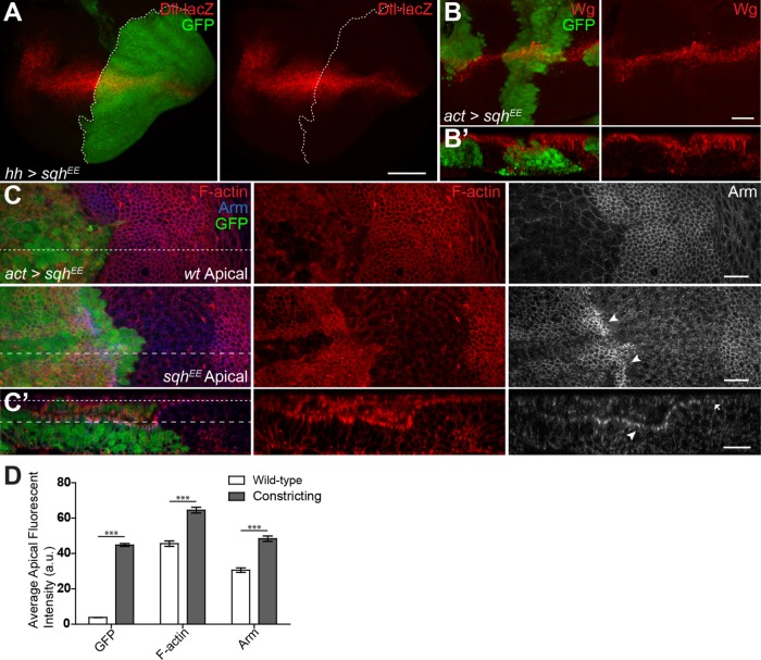
FIGURE 2:
NMII regulates Wg activity during wing development. (A) hh>sqhEE, gfp stained for Dll-lacZ expression. (B, B′) Total Wg in sqhEE flip-out clones, and cross-section showing cell constriction. (C, C′) GFP flip-out clones driving sqhEE stained for F-actin and Arm. (C′) Cross-section shows apical F-actin and Arm (C′, arrowhead vs. arrow) (dashed lines of C corresponding to section location and depth of C′). (D) Average fluorescence intensity of the apical surface of the wild-type and constricting columnar epithelial cells shown in C′. Data presented as mean ± SEM; ***p < 0.0001. Scale bars: (A) 50 µm, (B–C′) 20 µm.

